“Live reporter” technology has been demonstrated by synthetic biologists from Princeton University and Rice University to be able to reveal the functions of networks of signaling proteins in living cells with far greater precision than previous methods. The first-of-its-sort revealing device can show, for instance, how rapidly flagging organizations respond and how their reactions shift from one cell to another in both existences.
The tool was made by researchers using proteins that didn’t bother anyone and jumped on a crucial signaling mechanism that human cells use to control things like growth, differentiation, migration, inflammation, and other processes.
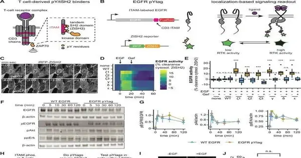
The Rice-Princeton team demonstrated their modular approach for tagging receptor tyrosine kinases (RTKs) with reporter proteins that activate green fluorescent proteins whenever their RTK partners become phosphorylated in the study, which was recently published in the open-access journal eLife.
Known as phosphorylation enzymes, kinases are enzymes that can alter the behavior of other proteins by attaching or detaching phosphate groups. RTKs are specialized kinases that, when they detect signals or stimuli from outside the cell, become phosphorylated and regulate essential cell functions.
The team demonstrated that the “live reporter” system could be utilized in conjunction with a microscope to produce a video record of the activity of signaling networks in living cells. According to Caleb Bashor, co-corresponding author of the study and assistant professor of both bioengineering and biosciences at Rice, the location and intensity of the signal network response can be determined by observing how brightly cells glow.
Play 00:00 00:03 Mute Settings PIP Enter fullscreen Play Timelapse of iRFP-ZtSH2 in NIH3T3 cells that co-express both EGFR-CD3-FusionRed and iRFP-ZtSH2. At the times indicated in the video, cells were first given gefitinib (10 M) before being given EGF (100 ng/mL). Credit: eLife (2023). DOI: 10.7554/eLife.82863
“More often than not, while you’re concentrating on stuff that occurs within cells, such as flagging organizations or quality organizations, you need to obliterate the cells to take a gander at their items,” Bashor said. “Whenever you can fabricate something where the phones stay alive and you can see how the flagging organization functions continuously within the cell, it’s an incredible benefit.”
In reference to biochemical nomenclature, in which tyrosine is referred to as “Y” and as “pY” when phosphorylated, the researchers gave the reporters the name pYtags.
Bashor and Xiaoyu Yang, a Ph.D. understudy in Bashor’s exploration bunch, created pYtags as a team with the examination gatherings of Princeton’s Jared Toettcher and Celeste Nelson. In human fibroblast cells, the study demonstrated that the system could record the activity of RTKs, or growth factor receptors.
According to Bashor, “We take an engineered protein that’s part of a different system—it’s actually part of immune signaling—and we put it into this new context, which is fibroblast cells that Jared works with in his lab at Princeton.” “We take an engineered protein that’s part of a different system.” Because it comes from a completely different kind of cell, we think it is probably not interacting with anything else in the cell. Therefore, it simply rests on the end of the growth factor receptor.
A proportional amount of fluorescence is produced when the pYtag reporter and its RTK partner co-activate. As a result, the cell glows brighter under a microscope, the stronger the RTK response.
Bashor stated, “It can receive that growth factor receptor phosphorylation signal.” As a result, the green fluorescent protein enters the cell when the receptor is activated, binds to something close to the membrane, and you see this green ring all around the cell’s exterior. That shows you when the cells are seeing the growth factor and how quickly the pathway is turning on in real time.
According to Bashor, pYtags can be used to monitor a variety of tyrosine kinase receptor types.
He stated, “We show in the paper that this reporter could be put onto multiple different types of growth-factor receptors, and that could be used as a reporter for all of them.” This provides a previously unattainable window into the dynamics of cellular signaling.”
More information: Payam E Farahani et al, pYtags enable spatiotemporal measurements of receptor tyrosine kinase signaling in living cells, eLife (2023). DOI: 10.7554/eLife.82863


