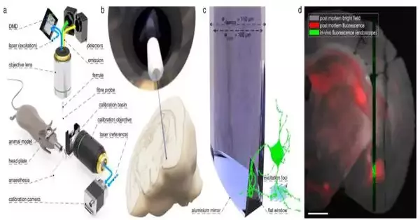
Minimally invasive techniques that allow for images of delicate deep-brain tissues are required to investigate the activity of neuronal structures and the interaction of nerve cells. An international team and Leibniz IPHT collaborated on the development of a new hair-thin endomicroscope that promises extremely gentle in-depth observations, the ability to investigate specific regions of the brain, and the onset and progression of severe neuronal diseases.
Neuroscientists hope that the instrument will assist them in developing novel approaches to combating these debilitating conditions. The findings were published by the researchers in Nature Communications.
Neuronal sicknesses like chemical imbalance, epilepsy, Alzheimer’s, or, alternately, Parkinson’s illness are still ineffectively perceived. As a result, preventing, treating, or curing these diseases remains a significant challenge. To all the more likely grasp the causes and the starting points of these sicknesses and to create and screen custom-made treatments, it is essential to translate and concentrate on what is meant for nerve cells, which are much of the time situated in exceptionally profound designs of the cerebrum, to act inside the regular intricacy of the entire organic entity.
“The endoscopic system is powered by an ultra-thin optical glass fiber that serves as a probe. We can photograph and observe individual cells and blood vessels in high-resolution, distortion-free, and high-contrast using digital holography. The hair-thin endoscopy design allows for extremely atraumatic in-vivo examination without causing damage to surrounding tissues,”
Prof. Dr. Tomáš Čižmár, head of the Fiber Research and Technology Research Department at Leibniz IPHT,
These conditions are studied by neuroscientists using small animal models and minimally invasive endoscopy to study deep-brain structures. To this end, researchers at the Leibniz Foundation of Photonic Innovation (Leibniz IPHT) in Jena, Germany, are cooperating with the group of mind-boggling Photonics from the Establishment of Logical Instruments of the Czech Institute of Sciences in Brno, Czech Republic, on a clever fiber-based approach.
Endoscope that is as thin as a human hair and is miniaturized The endoscope has a diameter of only 110 micrometers, making it possible to take pictures at subcellular and unprecedented depths of tissue. This not only makes it possible to investigate previously inaccessible deep-seated brain structures, but it also makes it possible to precisely investigate the neuronal connectivity and signaling activity of individual brain neurons.
“A probe made of an extremely thin optical glass fiber is at the center of the endoscopic system. We can use digital holography to image and visualize individual blood vessels and cells with high-contrast, distortion-free images.” Prof. Dr. Tomá Imár, head of the Fiber Research and Technology Research Department at Leibniz IPHT, who has led the development of the instrument, explains that the hair-thin endoscopy design enables extremely atraumatic in-vivo examination without damaging surrounding tissues.”
In in vivo neuroscience research, conventional endoscopic solutions typically make use of specialized rod lenses (GRIN), which transmit images from one end to the other. They may have a significant impact on the validity of neuroscience studies and a significant risk of tissue damage as a result of their footprint. By using a single multimode optical fiber as the imaging probe, the newly developed holographic endoscope overcomes this drawback and is the least invasive method for visualizing sensitive brain regions.
Additionally, the researchers have made use of a brand-new kind of fiber probe known as a side-view probe, which makes it possible to observe tissue that is perpendicular to the axis of the fiber. When compared to conventional bare-terminated probes, the endoscope’s insertion into the tissue causes much less stress and damage to the tissue being imaged. The side-view probe has the ability to go deeper into the tissue and can also stitch together individual frames taken along the way into a panorama-like image, giving a continuous view of the entire brain’s depth.
The developed endoscopic solutions permit the direct observation of intracellular processes as well as structural connectivity between neurons, particularly dendritic spines, which are microscopic structures that emerge from dendrites. It is possible to study the dynamics of subcellular structures, changes in intracellular calcium concentration (which are closely related to the signaling activity of nerve cells), and the speed of individual vessel blood flow. Neuroscientists may be able to identify signs of brain pathology based on any one of these properties.
“In neuronal diseases, changes or loss of nerve cells may limit the affected organism’s cognitive and motor performance forever. We are developing technologies for early detection of disease symptoms, such as altered neuronal communication. Prof. Dr. Tomá Imár says, “We can help to provide unprecedented insights into the control center of vital functions with high image quality and thus expand our understanding of neuronal diseases” with these new and powerful light-based instruments.
More information: Miroslav Stibůrek et al, 110 μm thin endo-microscope for deep-brain in vivo observations of neuronal connectivity, activity and blood flow dynamics, Nature Communications (2023). DOI: 10.1038/s41467-023-36889-z


