It has been generally acknowledged that tumorigenesis and disease movement comprise a multistep cycle. The most commonly involved strategy for disease finding and guessing to direct treatment choices depends on an intricate mix of imaging and obtrusive tissue biopsies. Nonetheless, the techniques are not generally delicate for beginning-phase disease finding.
Little extracellular vesicles (sEVs) are nanometer-sized, bilayer lipid transporters and contain a wide assortment of freight, including lipids, proteins, metabolites, RNAs, and DNA. sEVs let out by unique disease cells exist in practically all body liquids. They can turn into the likely flowing biomarkers in fluid biopsies, as they remarkably mirror the unique natural changes related to the developing growths and show the phases of disease movement.
The super-goal microscopy methods have arisen by pushing the goal as far as possible toward nanometer scales.
In another paper distributed in eLight, a group of researchers, driven by Teacher Dayong Jin from the College of Innovation Sydney, fostered a creative innovation in view of Lanthanide-doped EV-focusing on Nanoscopic Signal-speakers (Focal point). Their paper, “Upconversion Nanoparticles for Super-goal Evaluation of Single Little Extracellular Vesicles,” has huge potential in disease finding and guessing.
The kind of engineered upconversion nanoparticles (UCNPs) has non-direct photovoltaic switchable properties. They enable another kind of super-goal nanoscopy to accomplish sub-30 nm optical goals. The analyst’s new work involving nanophotonic tests additionally accomplished ultra-awareness in the quantitative location of sEVs. These tests recorded almost three significant degrees of awareness better than the standard protein-connected immunosorbent measure (ELISA).
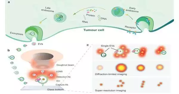
The imaging goal is further developed by the analysts to super-determine the surface biomarkers on single EVs (Fig. 1).The methodology depends on utilizing uniform, splendid, and photostable nanophotonic tests. Each is profoundly doped with a huge number of lanthanide particles.
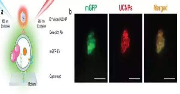
In their trial, the sEVs were first caught on a slide covered with CD9 immunizer and sandwiched by a biotinylated EpCAM neutralizer. Streptavidin functionalized upconversion nanoprobes, hence labeled the EpCAM immunizer for signal upgrade. The nanoprobes on single sEVs permit a super-goal magnifying lens for perception under a donut molded laser bar. A solitary nanoprobe in the donut bar creates an outflow design with a plunge where the test sits. Thus, the two close nanoprobes can be super-settled as far as possible at the nanoscale.

Fig. 2. (a) Co-limitation experiment diagram. (b) mGFP/UCNPs are labeled EpCAM+EVs when excited at 488 nm and 980 nm. Guan Huang, Yongtao Liu, Dejiang Wang, Ying Zhu, Shihui Wen, Juanfang Ruan, Dayong Jin
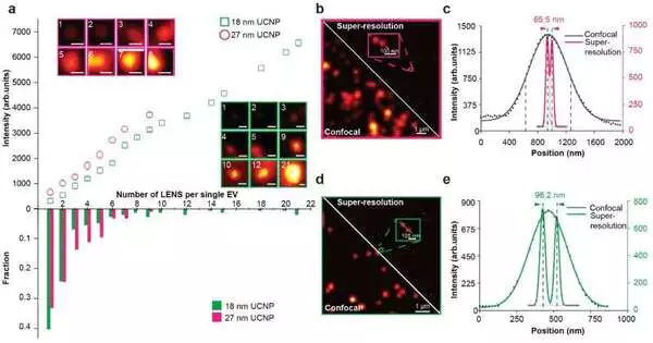
The scientists show that super-goal imaging of single sEVs can be accomplished utilizing a library of upconversion nanoprobes doped with different sorts of shifted groupings of producers. They affirm that immunizer formed nanoprobes can explicitly target growth epitope epithelial cell grip atom (EpCAM) on both huge EVs and single sEVs (Fig. 2). Utilizing super-goal imaging, the analysts can measure the particular number of nanoprobes on each sEV. They have shown that it is hypothetically conceivable to examine nanoprobes’ size and steric block on single sEVs (Fig. 3).

More information: Guan Huang et al, Upconversion nanoparticles for super-resolution quantification of single small extracellular vesicles, eLight (2022). DOI: 10.1186/s43593-022-00031-1


