Specialists have fostered an upgraded variant of optical rationality tomography (OCT) that can picture biomedical examples at higher differentiation and goal over a more extensive 3D field of view than was previously conceivable. The new 3D magnifying instrument could be helpful for biomedical exploration and, in the end, empower more precise clinical demonstrative imaging.
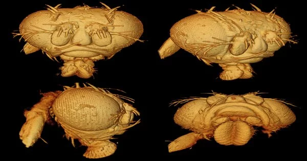
In the Optica diary, the analysts from Duke University describe the new procedure, which they call 3D optical cognizance refraction tomography (3D OCRT). Utilizing different organic examples, they show that 3D OCRT creates exceptionally point-by-point pictures that uncover highlights that are hard to see with conventional OCT.
OCT utilizes light to give high-quality 3D pictures without requiring any differentiation specialists or marks. In spite of the fact that it is regularly utilized for ophthalmology applications, the imaging strategy can likewise be utilized to picture numerous different pieces of the body, like the skin and inside the ears, the mouth, veins, and the gastrointestinal tract.
“The study reported in Optica expands on our prior research by overcoming substantial engineering obstacles, both in hardware and software, to enable OCRT to work in 3D and make it more generally applicable,” the researchers write.
Sina Farsiu research team co-leader
“OCT is a volumetric imaging method broadly utilized in ophthalmology and different parts of medication,” said first creator Kevin C. Zhou. “We fostered an intriguing expansion, including novel equipment joined with another computational 3D picture reproduction calculation to address a few notable limits of the imaging method.”
“We imagine this approach being applied in a wide assortment of biomedical imaging applications, like in vivo demonstrative imaging of the natural eye or skin,” said research group co-pioneer Joseph A. Izatt. “The equipment we intended to use to carry out the procedure can also be quickly scaled down into small tests or endoscopes to get to the gastrointestinal tract and other parts of the body.”

The new technique delivers exceptionally point-by-point pictures that uncover highlights hard to see with conventional OCT, as displayed in these pictures of a natural product fly head. Credit: Kevin Zhou, Duke University
Seeing more with OCT
Despite the fact that OCT has been demonstrated to be helpful both in clinical applications and biomedical examination, it is challenging to procure high-goal OCT pictures over a wide field of view every which way at the same time because of central constraints forced by optical bar proliferation. Another test is that OCT pictures contain elevated degrees of irregular commotion, called dots, which can obscure biomedically significant subtleties.
To address these impediments, the scientists utilized an optical plan that consolidated an illustrative mirror. This sort of mirror is generally found in non-imaging applications like electric lamps, where it encompasses the light to coordinate the light in one direction. The scientists involved an optical arrangement in which light was sent in the other direction, with the example of putting light where the light would be in a spotlight.
This plan made it conceivable to imagine the example from numerous perspectives over an extremely wide scope of points. They fostered a complex calculation to join the perspectives into a single excellent 3D picture that remedies for twists, commotion, and different defects.
“The work distributed in Optica reaches out on our past examination by defeating critical designing difficulties, both in the equipment and programming, to permit OCRT to work in 3D and make it all the more broadly relevant,” said research group co-pioneer Sina Farsiu. “Since our framework creates tens of gigabytes of information, we needed to foster another calculation in view of current computational apparatuses that have as of late developed inside the AI people group.”
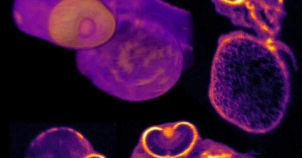
The video shows an examination between 3D OCRT and regular OCT renderings of a zebrafish hatchling. Credit: Kevin Zhou, Duke University
Getting a more extensive view
The scientists showed the flexibility and wide relevance of the strategy by utilizing it to model different natural examples, including a zebrafish and a natural product fly, which are significant model organic entities for conduct, formative, and neurobiological examinations. They likewise imaged mouse tissue tests of the windpipe and throat to show the potential for clinical analytic imaging. With 3D OCRT, they obtained 3D fields of perspective of up to 75° without moving the example.
“As well as decreasing clamor relics and adjusting for test-prompted twists, OCRT is innately prepared to do this computationally by making contrast from tissue properties that are less apparent in conventional OCT,” said Zhou. “For instance, we show that it is delicate to situate designs, for example, fiber-like tissue.”
The specialists are presently investigating ways of contracting the framework and making it quicker for live imaging by exploiting late improvements in quick OCT framework advancements and advances in profound discovering that can accelerate or further develop information handling.


