A high-level imaging-based strategy from researchers at Scripps Exploration offers a better approach for examining mitochondria.
In their report on February 14, 2023, in the Diary of Cell Science, the researchers portrayed a bunch of methods that empower the imaging and evaluation of even unobtrusive underlying changes inside mitochondria and the connection of those changes with different cycles progressing in cells.
Mitochondria are involved in energy creation and additionally in a few other basic cell capabilities, including cell division and cell-saving reactions to different sorts of pressure. Mitochondrial dysfunctions have been seen in a large group of illnesses, including Alzheimer’s, Parkinson’s disease, and various other diseases, and scientists are anxious to foster therapies that can switch these dysfunctions. However, the logical apparatus for concentrating on the finer nuances of mitochondrial structure has been restricted.
“We presently have a strong new toolbox for identifying and evaluating underlying, and in this manner utilitarian, contrasts in mitochondria—ffor instance, in sick versus solid states,” says concentrate on senior creator Danielle Grotjahn, Ph.D., a colleague teacher in the Branch of Integrative Primary and Computational Science at Scripps Exploration.
“We now have a potent new toolkit for detecting and quantifying structural, and consequently functional, changes in mitochondria—for example, in diseased versus healthy states.”
Senior author Danielle Grotjahn, Ph.D., assistant professor in the Department of Integrative Structural and Computational Biology.
The co-first creators of the review were Grotjahn Lab individuals Benjamin Barad, Ph.D., a postdoctoral exploration partner, and Michaela Medina, a Ph.D. competitor.
Mitochondria are one of the numerous film-bound atomic machines, or “organelles,” that abide inside the cells of plants and creatures. Regularly numbering in the hundreds to thousands for each phone, mitochondria have their own little genomes and have an unmistakable construction with an external layer and a wavy inward film where key biochemical responses happen. Researchers have discovered that the appearance of mitochondrial designs can change dramatically depending on what the mitochondrion is doing or what stresses are present in the phone.These primary changes in this manner can be exceptionally valuable markers of cell conditions; however, as of recently, there hasn’t been a decent technique for recognizing and evaluating them.
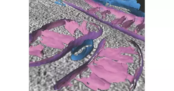
In another review, Scripps Exploration researchers portray new imaging procedures for examining mitochondria. Credit: Scripps Exploration
In the review, Grotjahn’s group set up a computational tool compartment to handle imaging information from a microscopy method called cryo-electron tomography (cryo-ET), which basically pictures organic examples in three aspects, utilizing electrons rather than light. The scientists’ “surface morphometrics tool compartment,” as they call it, empowers the nitty-gritty planning and estimation of the underlying components of individual mitochondria. This incorporates the twists of the internal film and the holes between layers—all possibly valuable markers of significant mitochondrial and cell events.
“It permits us basically to turn the delightful 3D pictures of mitochondria we can get from cryo-ET into delicate, quantitative estimations—wwhich we might possibly use to assist with distinguishing the point-by-point systems of illnesses, for instance,” Barad says.
The team used the toolbox to plan underlying subtleties on mitochondria when their cells were exposed to endoplasmic reticulum stress, a type of cell stress seen frequently in neurodegenerative diseases.They saw that key primary highlights like the shape of the inward film or the base distance among internal and external layers changed quantifiably when under this pressure.
The Grotjahn lab will now use their effective, rule-based exhibits of the new tool stash to focus on how mitochondria respond to cell stresses or other changes caused by illnesses, poisons, contaminations, and even drugs.
“We can look at the impacts on mitochondria in cells treated with a medication versus the consequences for untreated mitochondria, for instance,” Medina says. “What’s more, this approach isn’t restricted to mitochondria; we can likewise utilize it to concentrate on different organelles inside cells.”
“Measuring organellar ultrastructure in cryo-electron tomography utilizing a surface morphometrics pipeline” was co-authored by Benjamin Barad, Michaela Medina, Daniel Fuentes, Luke Wiseman, and Danielle Grotjahn, all of Scripps Exploration.
More information: Benjamin A. Barad et al, Quantifying organellar ultrastructure in cryo-electron tomography using a surface morphometrics pipeline, Journal of Cell Biology (2023). DOI: 10.1083/jcb.202204093


