
A study at the University of North Carolina School of Medicine published in the journal Science Advances examined brain processes implicated in neurological diseases. The researchers studied the impact of a neurotransmitter on brain network functional connectivity using fMRI and a genetic mouse model, a dynamic process important for human health and behavior. When you daydream, ruminate, consider the past, or plan for the future, a certain portion of your brain (in the prefrontal cortex) called the default mode network, or DMN, is shown to be most active.
The default mode network, or DMN, which includes part of the prefrontal cortex, is the area of your brain most active when you daydream, ruminate on something irritating, ponder the past, or prepare for the future. Scientists have long speculated that variations in DMN dynamics play important roles in some behaviors, such as those linked with attention deficit hyperactivity disorder, as well as diseases like Alzheimer’s and Parkinson’s, and conditions like depression and autism.
However, scientists do not fully comprehend the precise mechanisms that govern DMN dynamics. Researchers at the University of North Carolina School of Medicine, led by Ian Shih, Ph.D., associate professor of neurology, have now experimentally documented the interplay between neurons and brain chemicals across brain regions, resulting to changes in DMN dynamics.
Many brain imagers are very interested in finding the circuit processes that drive large-scale brain networks. However, how a specific neurotransmitter system impacts brain-wide dynamics is still unknown. Our findings serve to explain how norepinephrine influences brain activity and connectivity, resulting in DMN alterations.
Ian Shih
This study in mice, published in the journal Science Advances, provides evidence for how stimulating the locus coeruleus (LC), a tiny brain region in the brainstem that releases norepinephrine, alters DMN dynamics. It also proposes new therapeutic targets for restoring DMN function.
Early experiences have an impact on brain architecture development, which serves as the foundation for all future learning, behavior, and health. Just as a shaky foundation jeopardizes the quality and strength of a structure, poor childhood events can affect brain architecture, with long-term consequences.
“Many brain imagers are very interested in finding the circuit processes that drive large scale brain networks,” said Shih, senior author and head of the UNC Biomedical Research Imagine Center’s Center for Animal MRI (CAMRI) (BRIC). “However, how a specific neurotransmitter system impacts brain-wide dynamics is still unknown. Our findings serve to explain how norepinephrine influences brain activity and connectivity, resulting in DMN alterations.”
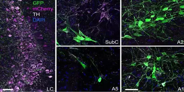
Shih and lead author Esteban Oyarzabal, Ph.D., a doctoral student at UNC-Chapel Hill at the time of the study, led functional magnetic resonance imaging (fMRI) investigations in a genetically engineered mouse model that expresses synthetic receptors in the LC. They then investigated the LC’s influence on the DMN.
The researchers were able to regulate brain cell activity by utilizing substances that selectively activate these synthetic receptors after developing a model to express these synthetic receptors. This “chemogenetic” method, developed by UNC pharmacology expert Byran Roth, MD, PhD, is ideal for Shih’s team’s fMRI manipulation of the LC. They discovered that stimulating the LC caused blood vessels in that region to contract while also enhancing low frequency fMRI activity changes in the DMN’s frontal cortical regions.
The researchers then developed an optical monitoring device that could simultaneously quantify the amount of norepinephrine released, neuronal calcium activity, and changes in brain blood volume. They demonstrated that norepinephrine from the LC can increase frontal cortical neuron spiking activity, while reducing blood volume.
“This has major implications for the interpretation of the fMRI data,” Shih said, “since it has been widely shown that neuronal and vascular activity in the brain are connected.” We now show that the presence of norepinephrine affects this connection.” They also revealed that chemogenetic activation of LC-NE neurons improved connectivity between neurons in the DMN’s frontal cortical regions. They discovered that the retrosplenial cortex and hippocampal regions of the brain can affect functional connectivity.
“We believe these two locations could serve as fresh targets to control frontal cortical regions and restore DMN function when LC neurons are deteriorated in Alzheimer’s and Parkinson’s disease,” Shih added.


