Attractive reverberation imaging (X-ray) machines can obviously see non-hard aspects of the body—ddelicate tissue like the cerebrum, muscles, and tendons—aas well as distinguish growths, making it conceivable to analyze numerous sicknesses and different circumstances. However, conventional MRI machines are only used in hospitals and other large facilities due to their high cost and bulk caused by their powerful magnets.
Companies are creating new portable versions with magnetic fields of lower strength as an alternative. The applications of MRI may be extended by these new models. For example, low-field X-ray frameworks could be sent in ambulances and other portable settings. They likewise could cost substantially less, encouraging X-rays to become all the more generally accessible, remembering underserved networks and agricultural countries.
However, more research is required to comprehend the relationship between low-field images and the underlying tissue properties they represent in order for low-field MRI scanners to reach their full potential. Scientists at the Public Foundation of Principles and Innovation (NIST) have been chipping away at a few fronts to propel low-field X-ray innovation and approve techniques for making pictures with more fragile and attractive fields.
“Magnetic resonance imaging of tissue varies with magnetic strength. The contrast of the pictures is different with low-field MRI equipment, thus we need to know how human tissue appears at these reduced field strengths.”
NIST electrical engineer Kalina Jordanova.
“Attractive reverberation pictures of tissue contrast are contingent upon attractive strength,” said NIST electrical specialist Kalina Jordanova. “Because the contrast of the images in low-field MRI systems is different, we must know how human tissue looks at these lower field strengths.”

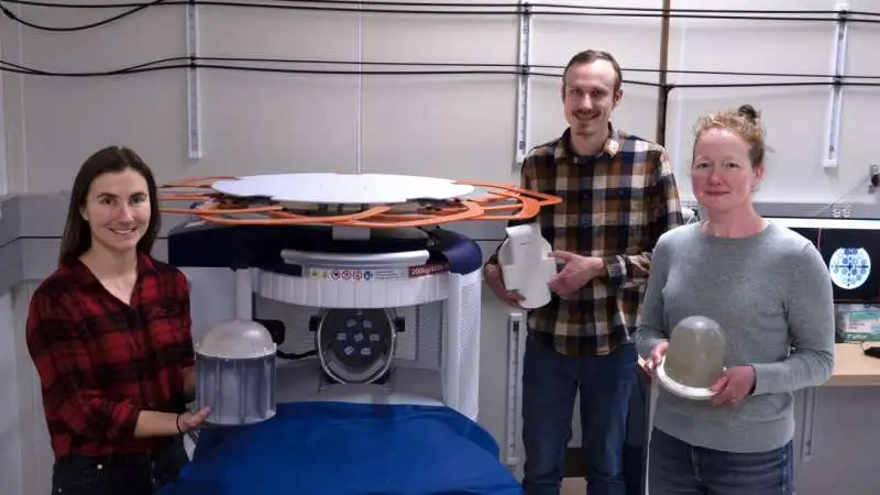
Among the NIST researchers who have been working on a number of projects, Kalina Jordanova, Stephen Ogier, and Katy Keenan are working to advance MRI technology that uses lower-strength magnetic fields and validate its methods for capturing images with weaker magnetic fields. Credit: R. Jacobson/NIST.
To accomplish this, researchers measured the properties of brain tissue with a weak magnetic field. Their outcomes were distributed in the diary Attractive Reverberation Materials in Physical Science, Science, and Medication.
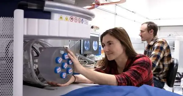
The specialists utilized a monetarily accessible compact X-ray machine to picture brain tissue in five male and five female workers. The pictures were made utilizing an attractive field strength of 64 millitesla, which is something like multiple times lower than the attractive field in customary X-ray scanners.
The team took pictures of the entire brain and gathered information about the gray matter, which contains a lot of nerve cells; the white matter, which is the deeper parts of the brain that contain nerve fibers; and the cerebrospinal fluid, which is the clear fluid that surrounds the brain and spinal cord.
“The MRI system is able to produce images that contain quantitative information about each constituent because these three brain constituents respond to the low magnetic field in distinct ways and produce distinct signals that reflect their distinct properties.” Knowing the quantitative properties of tissue permits us to foster new picture assortment techniques for this X-ray framework,” said NIST biomedical designer Katy Keenan.

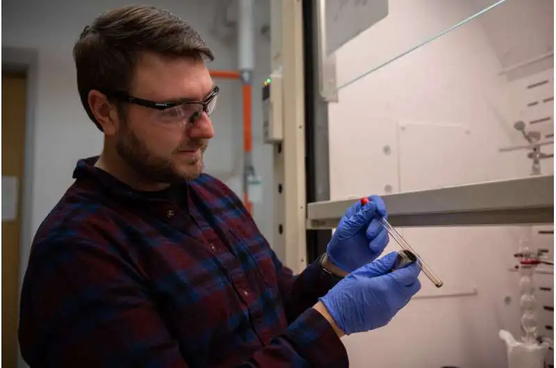
NIST scientist Sam Oberdick investigated contrast specialists for X-ray machines alongside his partner. His gathering tried iron oxide nanoparticles in lower-strength, attractive fields. These nanoparticles inside the fluid arrangement (imagined here) are attractive and are drawn toward the magnet through a mix of attractive connections and surface strain. Credit: R. Wilson/NIST.
In discrete work distributed in Logical Reports, NIST specialists teamed up with the College of Florence in Italy and Hyperfine Inc. in Guilford, Connecticut, to investigate a few up-and-comer materials that can essentially support picture quality in low-field X-ray filters.
At conventional magnetic field strengths, MRI contrast agents, which are magnetic materials that are injected into patients and enhance image contrast, make it simpler for radiologists to identify anatomical features or evidence of disease. However, researchers are only beginning to comprehend how the brand-new low-field MRI scanners might be used with contrast agents. At the lower field qualities of these scanners, contrast specialists might act uniquely in contrast to at higher field qualities, opening doors to involve new kinds of attractive materials for picture improvement.
In low magnetic fields, the sensitivity of various magnetic contrast agents was compared by NIST researchers and their colleagues. The specialists found that iron oxide nanoparticles outflanked conventional differentiation specialists, which are made of the component gadolinium, an intriguing earth metal. At low attractive field strengths, the nanoparticles gave great differentiation, utilizing a centralization of something like one-tenth that of the gadolinium particles.
Iron oxide nanoparticles likewise offer the benefit that they are separated by the human body rather than possibly aggregating in tissue, noted NIST scientist Samuel Oberdick. By correlation, a modest quantity of gadolinium might gather in tissue and could perplex the understanding of future X-ray checks in the event that it isn’t considered.
More information: Kalina V. Jordanova et al, In vivo quantitative MRI: T1 and T2 measurements of the human brain at 0.064 T, Magnetic Resonance Materials in Physics, Biology and Medicine (2023). DOI: 10.1007/s10334-023-01095-x
Samuel D. Oberdick et al, Iron oxide nanoparticles as positive T1 contrast agents for low-field magnetic resonance imaging at 64 mT, Scientific Reports (2023). DOI: 10.1038/s41598-023-38222-6


