Mass spectrometry imaging (MS imaging) gives exceptionally exact data on the spatial dispersion of substances in numerous areas. Scientists at the University of Bayreuth currently present model new applications in food examination in the journal Food Chemistry. Interestingly, they have prevailed with regards to making noticeable an added substance in dairy items and a creation-related tainting in prepared products. Extraordinary fixings that impact food quality can be recognized in natural products, vegetables, and meat items. The review, which was directed in collaboration with the Bavarian Health and Food Safety Authority (LGL), shows the extraordinary capability of this strategy, not least regarding purchaser assurance.
Natamycin in cheddar
To safeguard cheddar wheels or smoked hotdogs from mold pervasion, the surfaces are frequently treated with the fungicide natamycin. An EU guideline sets a boundary of one milligram for each square decimeter for this and furthermore specifies that natamycin should not infiltrate further than five millimeters into a treated cheddar wheel. This entrance profundity can’t be depicted exhaustively utilizing the food examination techniques regularly used to date, yet the Bayreuth research group driven by Prof. Dr. Andreas Römpp has had the option to utilize MS imaging to show interestingly where and in what amounts the fungicide happens in various kinds of Gouda.
“Our findings show that MS imaging is a beneficial supplement to existing food analysis methods, providing fresh insights into the geographical distribution and relative proportions of constituents. It has the significant advantage of not requiring the molecules of the ingredients to be labeled with dyes or other labeling methods. We will continue to work on refining the analytical capabilities of imaging mass spectrometry at the University of Bayreuth, within the newly established Faculty VII of Life Sciences: Food, Nutrition, and Health, in the future, combining it with other food analysis tools and applying it to previously unstudied ingredients. In this approach, we at Bayreuth University can make significant contributions to consumer protection.”
Prof. Römpp, who is the Chair of Bioanalytic Sciences and Food Analysis
The entrance of the natamycin atoms can be followed from the skin to within the cheddar wheel. The researchers worked together with the Bavarian Health and Food Safety Authority (LGL) on these examinations. In light of the outcomes acquired, they have created systemic principles for the recognizable proof of natamycin in cheddar. “Expanding on this recently evolved MS imaging approach, it could be feasible to diminish buyer openness to additives later on,” says Prof. Römpp, who is the Chair of Bioanalytic Sciences and Food Analysis at the University of Bayreuth.
Acrylamide in gingerbread
An EU guideline likewise draws certain lines for the presence of acrylamide in food. A malignant growth-advancing substance is shaped from sugar and asparagine—an amino corrosive—at low stickiness and temperatures over 120 degrees Celsius. A technique created in Bayreuth, Germany, in view of MS imaging, pictures acrylamide dispersion in customary German gingerbread. “To do this, we needed to cool the gingerbread tests to not exactly 60 degrees Celsius and then utilize an electric microsaw to create gingerbread cuts of two millimeter thickness. This was the main way we could distinguish tiny measures of acrylamide, “reports Prof. Römpp.”

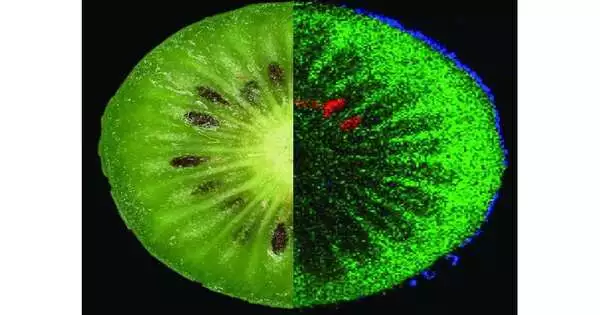
Elements of a kiwi: green = sugar, blue = polyphenol, and red = run of the mill kiwi lipid. Credit: Oliver Wittek.
Investigations of veal hotdogs
The new study likewise shows that MS imaging is similarly reasonable for investigations of handled meat items. In veal wieners, water-dissolvable and fat-dissolvable parts become apparent, so that low-fat and high-fat districts can be easily recognized. In like manner, it becomes apparent where substances of plant origin are tracked down that come from admixed spices. “However, MS imaging not just enables the limitation of fixings in meat items, but additionally helps, for example, in examinations of ‘tacky meat’ or something close by called hydrolysate added substances, which should pretend to be more excellent when they are not declared on the package.”It could consequently be valuable in distinguishing shopper trickery in meat items and better safeguard customers in this regard too, “says Prof. Römpp.
Kiwifruit and carrots
The application expected in the field of products of the soil is exhibited by concentrates on kiwifruit and carrots. The “smaller than expected kiwi” (Actinidia arguta) isn’t just sweet, but additionally has various wellbeing-advancing bioactive fixings. Utilizing test cuts that were a couple of hundredths of a millimeter thick and chilled off to a temperature of just 40 degrees, the Bayreuth bioanalysts pictured the circulation of a few substances in the skin and tissue: sugar particles (disaccharides), cell reinforcement polyphenol and a fat (lipid) normal for kiwis. In carrots, thus, particles of beta-carotene, a forerunner of vitamin A, were distinguished. It was also possible to distinguish the spatial distribution and normal sub-atomic designs of various colors (anthocyanins) that give carrots their orange, yellow, or violet hue.
A scientific technique without color.
“Our review clarifies that MS imaging is an important expansion to currently settled food investigation techniques. It offers new bits of knowledge into the spatial dispersion and relative extents of fixings. It enjoys the incredible benefit that the atoms of the fixings don’t need to be marked with colors or other naming strategies. At the University of Bayreuth—inside the recently settled Faculty VII of Life Sciences: Food, Nutrition, and Health—we will keep on working later on refining the logical abilities of imaging mass spectrometry, consolidating it with other food investigation apparatuses, and applying it to fixings not recently considered. “Along these lines, we at the University of Bayreuth can make significant commitments to customer security,” says Prof. Römpp.
On imaging mass spectrometry (MS),
For example, UV, fluorescence, infrared, or atomic attractive reverberation spectroscopy in that it isn’t subject to specific properties of the particles and iotas — for example, neither on light ingestion or fluorescence nor on atomic twist, the rakish force of a nuclear core around its focal point of gravity. On the off chance that two particles, or iotas, vary in mass, this distinction can be made noticeable by mass spectrometry. In this regard, a mass spectrometer is like a scale for iotas and particles—the main contrast being that it is a few million times more precise and delicate than any kitchen scale.
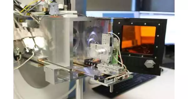
Before any mass spectrometric investigation, it is important to ionize the atoms of the substances to be recognized so that charged particles are made. This is on the grounds that charged particles can be diverted and advanced by the attractive and electric fields utilized in the mass spectrometer. Framework-aided laser desorption/ionization (MALDI) is one ionization technique used at the Chair of Bioanalytics and Food Analysis at the University of Bayreuth. Here, a grid substance is put on the example and then lit with a laser. Imaging mass spectrometry (MS imaging) consolidates data about particles got from MS with spatial data: by filtering an example surface and lighting an alternate spot on the example each time, pixel by pixel, a mass range can be recorded for each point that the laser has hit.
More information: Julia Kokesch-Himmelreich et al, MALDI mass spectrometry imaging: From constituents in fresh food to ingredients, contaminants and additives in processed food, Food Chemistry (2022). DOI: 10.1016/j.foodchem.2022.132529
Journal information: Food Chemistry


