Intrusive operations, for example, medical procedures requiring neighborhood sedation, frequently imply the risk of nerve injury. During activity, specialists may incidentally cut, stretch, or pack nerves, particularly while confusing them with another tissue. This can prompt enduring side effects in the patient, including tangible and engine issues. Also, patients getting nerve barricades or different sorts of sedation can experience the ill effects of nerve harm on the off chance that the needle isn’t set at the right distance from the designated fringe nerve.
Thusly, specialists have been attempting to foster clinical imaging strategies to moderate the risk of nerve harm. For example, ultrasound and attractive reverberation imaging (X-ray) can assist a specialist with pinpointing the area of the nerves during a procedure. Notwithstanding, it is trying to differentiate the nerves from the surrounding tissue in ultrasound pictures, while X-rays are costly and tedious.
In such a manner, there is a promising elective methodology known as multispectral photoacoustic imaging. A harmless strategy, photoacoustic imaging consolidates light and sound waves to make itemized pictures of tissues and designs in the body. Basically, the objective area is first enlightened with beat light, making it heat up somewhat. This, thus, makes the tissues grow, conveying ultrasonic waves that can be detected by an ultrasound finder.
“Our study is the first to characterize the optical absorbance spectra of fresh swine nerve samples using a broad spectrum of wavelengths, as well as the first to show in-vivo visualization of healthy and regenerated swine nerves with multispectral photoacoustic imaging in the NIR-III window.”
Dr. Muyinatu A. Lediju Bell,
As of late, an exploration group from Johns Hopkins College led a concentrated study in which they completely portrayed the retention and photoacoustic profiles of nerve tissue across the near-infrared (NIR) range. Their work, distributed in the Diary of Biomedical Optics, was driven by Dr. Muyinatu A. Lediju Ringer, John C. Malone Academic Administrator and Heartbeat Lab Chief at Johns Hopkins College.
One of the principal targets of their review was to determine the best frequencies for recognizing nerve tissue in photoacoustic pictures. The specialists conjectured that the frequencies from 1630–1850 nm, which dwell inside the NIR-III optical window, would be the ideal range for nerve representation since the lipids found in the myelin sheath of neurons have a trademark retention top here.
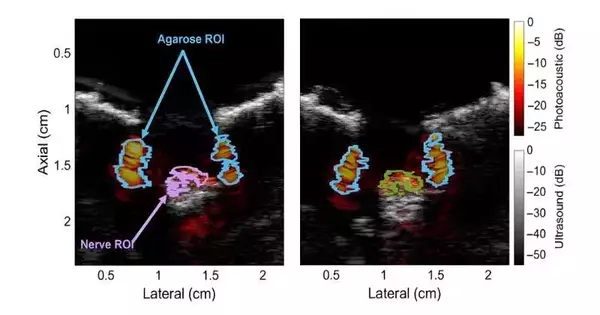
To test this speculation, they performed point-by-point optical ingestion estimations on fringe nerve tests acquired from pigs. They noticed an absorbance peak at 1210 nm, which fell in the NIR-II range. Notwithstanding, such an assimilation top is additionally present in different kinds of lipids. Conversely, when the contribution of water was deducted from the absorbance range, nerve tissue displayed a one-of-a kind peak at 1725 nm in the NIR-III reach.
Furthermore, the specialists conducted photoacoustic estimations on the fringe nerves of live pigs utilizing a custom imaging arrangement. These tests additionally affirmed the speculation that the top of the NIR-III band can really be utilized to separate lipid-rich nerve tissue from different kinds of tissues and materials containing water or that are lipid-lacking.
Happy with the outcomes, Ringer comments, “Our work is quick to describe the optical absorbance spectra of new pig nerve tests utilizing a wide range of frequencies, as well as the first to exhibit in-vivo perception of sound and recovered pig nerves with multispectral photoacoustic imaging in the NIR-III window.”
In general, these discoveries could spur researchers to additionally investigate the capabilities of photoacoustic imaging. Also, the portrayal of the optical absorbance profile of nerve tissue could assist with further developing nerve discovery and division procedures while utilizing other optical imaging modalities.
“Our outcomes feature the clinical commitment of multispectral photoacoustic imaging as an intraoperative method for deciding the presence of myelinated nerves or forestalling nerve injury during clinical intercessions, with potential ramifications for different optics-based innovations. Our commitments subsequently effectively lay out another logical starting point for the biomedical optics local area,” closes Ringer.
More information: Michelle T. Graham et al, Optical absorption spectra and corresponding in vivo photoacoustic visualization of exposed peripheral nerves, Journal of Biomedical Optics (2023). DOI: 10.1117/1.JBO.28.9.097001


