A fundamental job of the cornea is to focus light on the retina. Its bended shape, and consequently its capacity to shine light, should be completely steady to guarantee a sharp picture in spite of the different shocks and rubs it gets throughout the day. The cornea is subsequently made out of a few hundred collagen lamellae stacked one on top of the other with different in-plane directions.
Yet, the straightforwardness of this tissue, fundamental for visual keenness, is an impediment to optical imaging because of its design, which utilizes the customary optical magnifying lens. Subsequently, the unfortunate portrayal of the lamellar construction of the cornea today restricts how we might interpret the connection between the design and capability of this tissue, especially with regards to its mechanical properties. Another restriction concerns the comprehension of specific pathologies connected to faulty construction, like keratoconus.
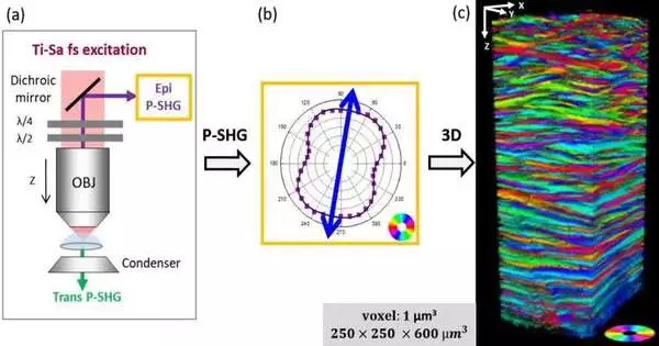
In another paper distributed in Light: Science and Applications, a group of researchers, led by Teacher Marie-Claire Schanne-Klein from the Lab for Optics and Biosciences at Ecole Polytechnique, CNRS, Inserm, and Institut Polytechnique de Paris, Palaiseau, France, have utilized a new three-layered (3D) optical imaging procedure: second symphonious age (SHG) microscopy, which empowers explicit representation of collagen with next to no earlier naming.
The creativity of their methodology lies in playing on the polarization of light to uncover the heading of the collagen fibrils that make the lamellae out of the cornea. This imaging is performed, all things being equal, utilizing epi-location and might additionally evolve to empower in vivo analysis ultimately. One more benefit of this setup is that it gives undeniably more exact outcomes than transmission.
This is on the grounds that the soundness length of SHG processes is more limited in the retrogressive bearing than in the forward heading, which by and large guarantees a better spatial goal. Ten unblemished human corneas were described in this way. A cautious approval was performed by two self-referred-to strategies, as SHG polarimetry had never been carried out on tissue of such thickness (around 600 m).
Specifically, the creators have checked that a similar construction is obtained by imaging the cornea in two distinct directions. This study has accordingly displayed interestingly that, while the lamellae of the human cornea are worldwide situated in two opposite bearings, their fundamental course changes with profundity.
This study prepares for promising new portrayals of the cornea: planning the size and dissemination of lamellae as an element of profundity, yet additionally as a component of position (focus versus outskirts of the tissue). This data will take care of the mechanical demonstration of corneal conductivity because of variations in intraocular strain or mending processes. At long last, the investigation of obsessive tissues will empower us to lay out the job of corneal design in specific illnesses.
More information: Clothilde Raoux et al, Unveiling the lamellar structure of the human cornea over its full thickness using polarization-resolved SHG microscopy, Light: Science & Applications (2023). DOI: 10.1038/s41377-023-01224-0


