Materials researchers are frequently bioinspired, and in another review, bird-propelled by primary varieties shown by avian species to frame non-luminous nanoparticle gatherings. In order to identify structure-color relationships and produce designer materials with individualized color, such nanoparticle mixtures with varying particle chemistry and size can affect the color that is produced.
Christian M. Heil and a group of researchers from a number of international, multidisciplinary research institutes in the United States, Belgium, and Germany demonstrated how to use small-angle scattering measurements to reassemble the structures in a new Science Advances article.
In order to demonstrate the influence of a single layer of segregated nanoparticles and produce a color of interest, the research team was able to accurately and quantitatively predict the colors that were observed in the experiments in mixtures with strongly absorbed nanoparticles. It was possible to engineer synthetic materials with the desired colors for use in paints, cosmetics, and food coloring thanks to the adaptable computational methods that were incorporated into this work.
Fabrication and characterization of synthetic colors
The numerous hues found in nature serve as inspiration for the color fabrication of synthetic materials. They may originate from the spatial organization of color-resistant nanostructured materials. Colors that are iridescent can be produced by materials with consistent, periodic nanostructures, whereas colors that are not iridescent are produced by materials with short-range ordering.
By self-assembling polymeric nanoparticles through amorphous assemblies of inorganic nanoparticles, material scientists can imitate natural structural colors that are not iridescent. They can vary the shades of nanoparticle congregations by managing their construction and optical properties to expand their underlying variety. By generating a wide variety of distinct structures and structural colors, researchers are able to adjust the ratio of the nanoparticle size to the size of the components and the composition.
Heil et al.’s description of designer materials used electron microscopy and small-angle scattering to obtain structural information about the designer materials. They added small-angle neutron and X-ray scattering integration to offer suitable approaches for analyzing information about the structure at the nanoscale and in bulk. For optical demonstration of complex nano-congregations, they utilized the limited distinction time-space (FDTD) strategy.
They used computational reverse-engineering analysis for scattering experiments (CREASE) to interpret the small-angle scattering experiments’ profile to generate nanoparticle assemblies.
The specialists used wrinkle FDTD techniques to comprehend paired nanoparticle gatherings framing supraballs to create a wide range of varieties. The results showed that nanoparticle assemblies’ optical properties could be customized for a wide range of applications in food coloring, cosmetics, and paint.
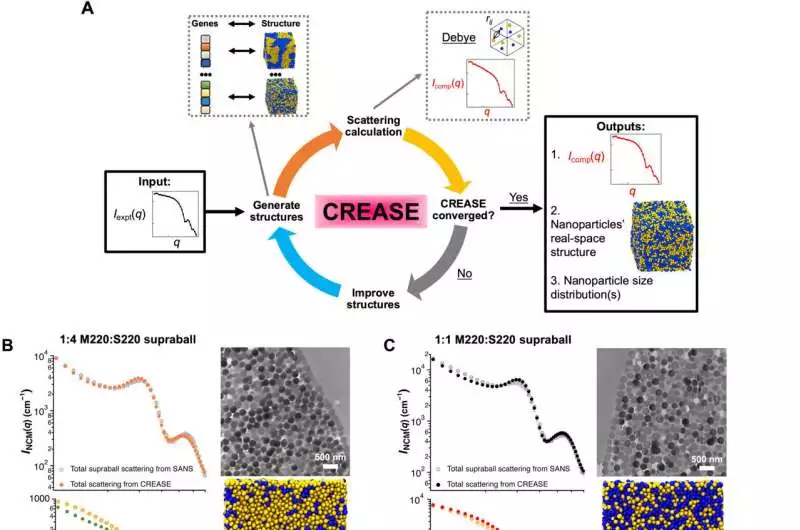
Applying the wrinkle strategy to recreate the paired nanoparticle combination gathering structure from SANS profiles (A) Schematic showing how the CREASE method works B to D) I as a function of q for NCM in a SANS plot (left top; gray dots) as well as MCM (bottom left; overlaid with the scattering profile of the CREASE output structures for NCM and MCM conditions for (B) 1:4, (C) 1:1, and (D) 4:1 melanin (yellow dots): silica-based products. B) plots CREASE’s NCM and MCM as orange and green, respectively, with a 2 scattering error of 2.35; (C) colors CREASE’s NCM and MCM as black and red, respectively, with a 2 scattering error of 2.03; and (D) displays CREASE’s NCM and MCM as blue and purple, respectively, with a 2 scattering error of 2.01. On the right, the transmission electron micrograph of the cross section of a typical binary mixture supraball (right top) is shown in B to D. spheres of lighter melanin; silica, more obscure circles; scale bars, 500 nm); and a Visual Molecular Dynamics (VMD) visualization of the central portion of the reconstructed 3D binary nanoparticle mixture assembly, which measures three millimeters by three millimeters by ten millimeters. Yellow spheres represent silica chemistry, and blue spheres represent melanin (right bottom). C) The scattering profile and CREASE are based on (50). E) Scanning electron micrographs of a representative supraball and supraball surface from a 1:4 binary mixture (left to top; scale bars are 100 nm and 1 m, respectively) and the lognormal size distribution of the melanin nanoparticles that were used to create the surface-segregated shell following CREASE reconstruction (left bottom). VMD representation of the focal piece (3 m by 3 m by 10 m) of the reproduced 3D twofold nanoparticle combination gets together with the additional melanin shell layer (right). Credit: Science Advances, doi: 10.1126/sciadv.adf2859
Structural color diversity with binary nanoparticle mixtures
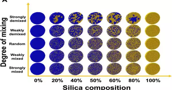
Materials scientists looked at the phase morphology and composition of binary mixtures of absorbing and non-absorbing species of similar size to find a way to control the prediction of structural colors that were used to determine structural color’s relative sensitivity. This work produced a wide range of colors to demonstrate the necessity of knowing the relative composition, phase segregation, and mixing extent of two distinct types of nanoparticles in a binary mixture.
To screen this cycle, Heil et al. used a combination of experimental and computational methods to predict each reflectance spectrum’s color and the internal morphologies of supraballs.
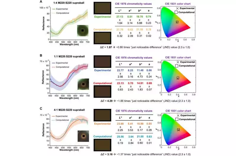
Optical display and variety investigation examination between trial supraballs and FDTD computations on the wrinkle yield structures (A) Reflectance spectra (experimental, solid curve; run bend, figured), RGB variety board, CIE 1976 chromaticity values, and CIE 1931 chromaticity directions’ examinations for the 1:4 melanin: supraball system binary mixture of silica A color difference (E) value that is less than 0.9 times the average JND value can be used to calculate the quantitative difference between the FDTD and experimental colors. The area (3 m by 3 m) that was probed during optical measurements using a microspectrophotometer is depicted by the black box in the inset of the optical micrograph of the corresponding supraball. B) Similar to (A), but with one-to-one melanin: silica double blend supraball framework with an E esteem that is 1.9 times the typical JND esteem. (C) Much like (A), but with a 4:1 melanin: supraball system of a binary mixture of silica with an E value that is 1.4 times greater than the typical JND value. The standard deviation (SD) is depicted in the reflectance plots by the lighter-colored envelopes that result from the experimental and computational lines. The SD of the computational methodology is from FDTD reproductions of three freely produced wrinkle structures, and the SD of the exploratory estimations is from 15 autonomous estimations of various supraballs. Credit: Science Advances, doi: 10.1126/sciadv.adf2859.
Data from CREASE and small-angle neutron scattering
The team produced suspensions of micron-scale supraballs and carried out small-angle neutron scattering experiments on three distinct nanoparticle mixtures. In order to collect total scattering from the nanoparticles of interest, they isolated the scattering contribution of individual components within the structure to obtain morphological information about the interior of supraballs.
Involving computational figuring out examination for the dissipating tests, the group investigated dispersing results from multicomponent designs to acquire 3D underlying recreation and optical reproductions of the items.
The coordinates of all nanoparticles within supraballs were provided by the results of the structural analysis based on computational reverse-engineering analysis for scattering experiments (CREASE). The finite-difference-time domain method (FDTD) used this information to calculate light scattering.
With optical modeling comparisons, the combined CREASE-FDTD method represented a structural match that was very close to the experimental systems. The team modeled the optical properties underlying a complex binary mixture of nanoparticles in order to comprehend the effect of size dispersion and surface segregation on structural color. This was done in light of the remarkable agreement between these two combined methods.
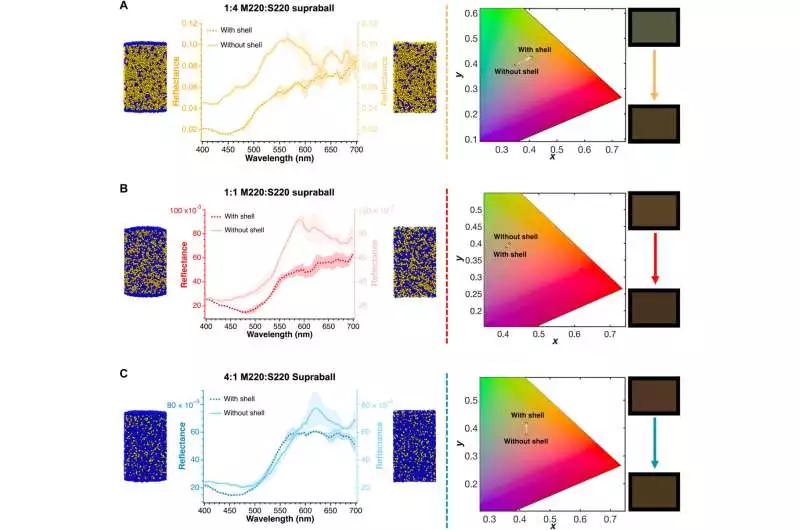
Comparative color analysis and optical modeling of the CREASE output structures calculated using FDTD with and without the melanin shell on the structures. A-C) Reflectance spectra (a solid curve containing a melanin shell; run bend, without melanin shell), structure representations, CIE 1931 chromaticity directions’ examinations, and RGB variety board examinations for the (A) 1:4, (B) 1:1, and (C) 4:1 melanin: supraball systems binary mixture of silica. The lighter hued envelopes coming from with and without shell processed reflectance profiles in the reflectance plots demonstrate the SD. For each instance, the SD is derived from FDTD simulations of three independently generated CREASE structures. Credit: Science Advances, doi: 10.1126/sciadv.adf2859
Outlook
In this manner, Christian M. Heil and coworkers combined two important techniques: Using the finite-difference-time domain method (FDTD) and computational reverse engineering analysis for scattering experiments (CREASE), a new platform has been created to model the structural colors of nanoparticle-based supra-assemblies. Non-iridescent nanoparticle assemblies were formed by structural colors inspired by birds, while colors with short-range ordering are also produced.
The results as a function of the nanoparticles’ size, dispersion, phase morphology, and strongly absorbing optical properties were determined by the team using two primary methods. This joined strategy created reproduced 3D designs of nanoparticle gatherings with dissipating profiles that were a near match to those obtained through little point neutron dispersing estimations. For the purpose of studying the optical properties of much larger assemblies of supraballs, such as packed films and pigment dispersions, the combined approach is well-suited for multiscale modeling.
This method can be used to create programmable colors that can be used in cosmetics, coatings, and paints. It can also help researchers in materials better understand and design complex materials for applications across the electromagnetic spectrum. It can also predict material properties like thermal, electrical, and mechanical properties, which are affected by the materials’ different structural compositions.
More information: Christian M. Heil et al, Mechanism of structural colors in binary mixtures of nanoparticle-based supraballs, Science Advances (2023). DOI: 10.1126/sciadv.adf2859
Vukusic P. et al, Photonic structures in biology. Nature (2023), DOI: 10.1038/nature0194


