As a tool for improving illness detection, diagnosis, and clinical care, artificial intelligence is set to change the profession of radiology. The device has the potential to help clinicians by revealing hidden information in imaging scans that is undetectable to the naked eye.
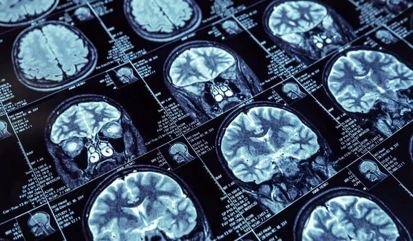
Columbia University researchers show in an article published in JAMA Oncology that applying artificial intelligence to standard-of-care imaging may help predict how well immunotherapy will work for patients with melanoma (link is external and opens in a new window).They created a machine learning algorithm that analyzes a patient’s computed tomography (CT) scans and generates a biomarker called a radiomic signature that corresponds with the patient’s outcome.
The signature uses certain aspects of the tumor to predict whether a patient’s disease will respond well to immunotherapy, remain stable, or progress with high accuracy. Immunotherapy, which has become a popular melanoma treatment, works by stimulating a patient’s own immune system to fight cancer.
“We hope to take a patient early on who looks like they are not doing well on a given therapy because of their signature and enhance, change, or add another drug to the therapy,”
Lawrence H. Schwartz
“We hope to take a patient early on who appears to be failing on a given therapy due to their signature and enhance, change, or add another drug to the therapy,” says Lawrence H. Schwartz, MD, James Picker Professor and chair of the Department of Radiology at Columbia University Vagelos College of Physicians and Surgeons (VP&S). “[The goal is to] truly optimize customized cancer care in real time for each patient.”
Dr. Schwartz, a member of the Herbert Irving Comprehensive Cancer Center (HICCC), hopes to broaden the experiment with his colleagues to include other tumor types, such as lung cancer, colon cancer, kidney cancer, and prostate cancer, as well as treatments other than immunotherapy. The researchers sought to start with a novel therapy and picked melanoma because of the disease’s recent, rapid uptake of immunotherapy.
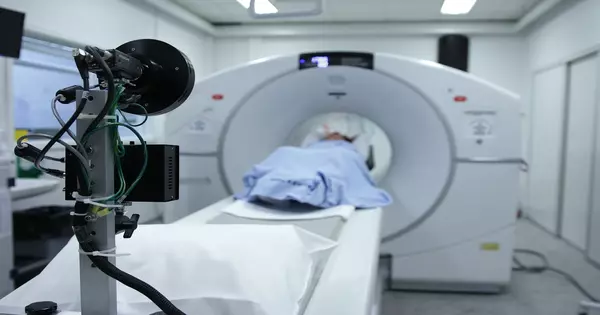
Currently, clinicians judge the value of a therapy almost entirely based on tumor size. After therapy begins, patients have a baseline CT scan followed by periodic follow-up scans. If the tumor decreases, the treatment appears to be effective, whereas growth indicates that the patient’s illness is worsening.
However, with immunotherapy, this is not always the case, as studies have shown that tumor size and growth do not always correspond with overall survival.
Most of the existing response criteria were designed some decades ago to measure the response to systemic treatments like chemotherapy, explains Laurent Dercle, MD, PhD, an associate research scientist in the Department of Radiology at VP&S. “Immunotherapy has created new patterns of response and progression, with some patients experiencing a temporary rise in tumor growth followed by a response.” As a result, we needed to develop new measures to predict therapy success. “
Tumors may evolve biologically throughout the course of a patient’s condition in ways that are more complex than a simple measurement of size can indicate. As an example, the researchers discovered that their machine learning algorithm performed best when it took into account not only tumor volume and growth, but also tumor spatial heterogeneity, or the non-uniform distribution of cancer cells across disease sites, and texture, which examines the variation of pixel intensities across the tumor CT image.
The algorithm was verified using data from 287 patients with advanced melanoma who took part in the multicenter clinical trials KEYNOTE-002 (link is external and opens in a new window) and KEYNOTE-006 (link is external and opens in a new window) that used the immunotherapy medication pembrolizumab. The radiomic signature, which used baseline and 3-month follow-up CT images, was able to predict overall survival at 6 months with a high degree of accuracy. Indeed, it surpassed the usual method based on tumor diameter, known as Response Evaluation Criteria in Solid Tumors 1.1 (RECIST 1.1), which is routinely used in clinical studies to evaluate treatment efficacy.
“With this artificial intelligence revolution, the area of radiology and imaging in general has never been more interesting,” adds Dr. Schwartz. “We’ve always looked at advancements in terms of new equipment, new tracers, and other such things.” However, this provides us with an opportunity to leverage the information we have from all of our imaging modalities in order to accelerate diagnosis, improve accuracy and precision, and provide patients with more effective treatments.”
Additional Information:
“Early Readout on Overall Survival of Patients With Melanoma Treated With Immunotherapy Using a Novel Imaging Analysis” is the title of the paper. All the
authors are from Columbia University: Laurent Dercle (University of Columbia), Binsheng Zhao (University of Columbia), Mithat Gönen (MSKCC), Chaya S. Moskowitz (MSKCC), Ahmed Firas (University of Columbia), Volkan Beylergil (University of Columbia), Dana E. Connors (National Institutes of Health), Hao Yang (University of Columbia), Lin Lu (University of Columbia), Tito Fojo (University of Columbia).Richard Carvajal (Columbia), Sanja Karovic (Inova Center for Personalized Health and Schar Cancer Institute), Michael L. Maitland (University of Virginia Cancer Center), Gregory V. Goldmacher (Merck & Co, Inc.), Geoffrey R. Oxnard (Dana Farber Cancer Institute), Michael A. Postow (MSKCC and Weill Cornell), and Lawrence H. Schwartz (Columbia).


