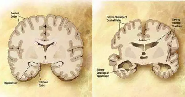
One of the main features of Alzheimer’s disease is neurodegeneration, or the gradual loss of neuron function. However, not all brain regions are affected equally.
One of the principal cerebrum locales to show neurodegeneration in Alzheimer’s disease is a piece of the nerve center called the mammillary body. A subset of neurons in this body that are most susceptible to neurodegeneration and hyperactivity have been identified by MIT researchers in a new study. Additionally, they discovered that this damage impairs memory.
According to the researchers, the findings suggest that this region may contribute to some of the earliest Alzheimer’s disease symptoms, making it a suitable target for potential new drugs to treat the disease.
Li-Huei Tsai, senior author of the study and director of MIT’s Picower Institute for Learning and Memory, says, “It is fascinating that only the lateral mammillary body neurons, not those in the medial mammillary body, become hyperactive and undergo neurodegeneration in Alzheimer’s disease.”
“It is intriguing that only the lateral mammillary body neurons, not those in the medial mammillary body, experience neurodegeneration and hyperactivity in Alzheimer’s disease.”
Li-Huei Tsai, director of MIT’s Picower Institute for Learning and Memory and the senior author of the study.
By treating mice with a drug that is currently used to treat epilepsy, the researchers demonstrated that they could reverse memory impairments caused by hyperactivity and neurodegeneration in mammary body neurons.
The lead authors of the paper, which was published today in Science Translational Medicine, are Zhuyu (Verna) Peng and Mitchell Murdock, graduate students at MIT, and Wen-Chin (Brian) Huang, a former postdoctoral researcher at MIT.
Predisposed to degeneration
As Alzheimer’s disease progresses, neurodegeneration and the formation of tangles in the brain by misfolded tau proteins and amyloid beta plaques also occur. One unanswered question is whether this neurodegeneration affects all neurons equally or whether some types of neurons are more susceptible.
Murdock asserts, “We would have a better understanding of neurodegeneration if we could identify specific molecular properties of classes of neurons that are predisposed to dysfunction and degeneration.” This is clinically significant on the grounds that we could track down approaches to remedially focus on these weak populations and possibly defer the beginning of mental deterioration.”
Tsai, Huang, and others found that the mammillary bodies—a pair of structures on the left and right undersides of the hypothalamus—had the highest density of amyloid beta in a 2019 study using a mouse model of Alzheimer’s disease. Although their precise function in both normal memory and Alzheimer’s disease is unknown, it is known that these bodies are involved in memory.
The researchers used single-cell RNA-sequencing, which can reveal the genes that are active within various types of cells in a tissue sample, to learn more about the function of the maternal body. The researchers found two major neuronal populations by employing this strategy: one in the medial and one in the lateral parts of the mammary bodies. The researchers found that synaptic activity-related genes were highly expressed in the lateral neurons, and these neurons also spiked more frequently than the medial mammalian body neurons.
In light of those distinctions, the analysts contemplated whether the parallel neurons may be more defenseless to Alzheimer’s illness. To investigate that inquiry, they concentrated on a mouse model with five hereditary changes connected to the beginning stages of Alzheimer’s in people. The analysts found that these mice showed substantially more hyperactivity in horizontal mammillary body neurons than sound mice. In any case, the average mammary body neurons in solid mice and the Alzheimer’s model showed no such contrasts.
The researchers discovered that this hyperactivity began before amyloid plaques began to form, around two months of age, which is equivalent to a young adult. As the mice got older, the lateral neurons became even more hyperactive, and they were also more likely to die than the medial neurons.
According to Murdock’s assertion, “We think the hyperactivity is related to dysfunction in memory circuits and is also related to a cellular progression that may lead to neuronal death.”
The Alzheimer’s mouse model showed impedances in framing new recollections, yet when the specialists treated the mice with a medication that decreases neuronal hyperactivity, their presentation on memory errands was essentially taken to the next level. Levetiracetam is a medication that is used to treat epileptic seizures. It is also being tested in clinical trials to treat epileptiform activity, which is hyperexcitability in the cortex that increases the risk of seizures, in Alzheimer’s patients.
Contrasting mice and people
The specialists likewise concentrated on human cerebrum tissue from the Strict Orders Study/Memory and Maturing Undertaking (ROSMAP), a longitudinal report that has followed memory, engine, and other age-related issues in more established individuals starting around 1994. The researchers found two clusters of neurons that correspond to the lateral and medial mammary body neurons they found in mice by using single-cell RNA-sequencing of mammary body tissue from people with and without Alzheimer’s disease.
Overexpression of genes encoding potassium and sodium channels was also found in the lateral mammillary bodies of Alzheimer’s tissue samples, which the researchers compared to the mouse studies. They also found that the lateral neuron cluster had more neurodegeneration than the medial cluster in those samples.
Early in the disease, other studies of Alzheimer’s patients have found that the mammary body loses volume, plaques build up, and synaptic structure changes. According to the researchers, all of these findings suggest that the maternal body might be a good target for drugs that could halt the progression of Alzheimer’s disease.
Tsai’s lab is currently chipping away at additional characterization of how the parallel neurons of the mammillary body are associated with different pieces of the mind to sort out how it structures memory circuits. Additionally, the researchers hope to learn more about the characteristics of the mammalian body’s lateral neurons that make them more susceptible to neurodegeneration and amyloid deposition.
More information: Wen-Chin Huang et al, Lateral mammillary body neurons in mouse brain are disproportionately vulnerable in Alzheimer’s disease, Science Translational Medicine (2023). DOI: 10.1126/scitranslmed.abq1019. www.science.org/doi/10.1126/scitranslmed.abq1019


