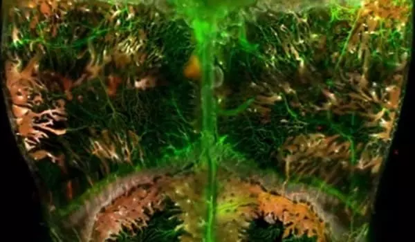
Scientists created a 3D map of the blood vessels and self-renewing ‘stem’ cells that line and penetrate a mouse skull using glowing chemicals and other techniques. The map pinpoints the locations of blood vessels and stem cells, which scientists hope to use to repair wounds and regenerate bone and tissue in the skull.
Scientists at Johns Hopkins Medicine used glowing chemicals and other techniques to create a 3D map of the blood vessels and self-renewing “stem” cells that line and penetrate the skull of a mouse. The map pinpoints the locations of blood vessels and stem cells, which scientists hope to use to repair wounds and regenerate bone and tissue in the skull.
“We need to see what’s going on inside the skull, including the relative locations of blood vessels and cells and how their organization changes during injury and over time,” says Warren Grayson, Ph.D., a biomedical engineering professor and director of the Laboratory for Craniofacial and Orthopaedic Tissue Engineering at Johns Hopkins University School of Medicine. His research focuses on the development of biomaterials and the transplantation of stem cells into the skull to replace missing bone tissue.
We need to see what’s going on inside the skull, including the relative locations of blood vessels and cells and how their organization changes during injury and over time. A larger image of the skull gives us a better understanding of the entire vasculature and distribution of different stem cell types.
Warren Grayson
Other researchers have created maps of blood vessels and stem cells in the mouse skull. “However, a larger image of the skull gives us a better understanding of the entire vasculature and distribution of different stem cell types,” says Alexandra Rindone, first author of the paper and graduate student at The Johns Hopkins University and School of Medicine.
The new map, published in Nature Communications, is a 3D view of the top of a mouse skull its cranial bone, or calvaria which is made up of four connected skull bones.
The Johns Hopkins researchers used four key techniques to pinpoint vessels and cells to create the map, which includes hundreds of thousands of cells. They began by using immunofluorescence to label molecules on the surface of various blood vessels and stem cells with a fluorescent or glowing, chemical. The scientists then use a chemical compound that allows light to pass through the skull without scattering – a technique known as optical tissue clearing. “It makes the skull look like glass,” Rindone says.
The scientists used a light-sheet microscope to capture the 3D image, which captures images of large sections of tissue at high resolution and speed while minimizing photo bleaching. “This tool assists us in avoiding deterioration of the fluorescent dye when tissues are exposed to light sources for an extended period of time,” Rindone says.
Finally, they used computer software to identify and segment the skull’s 3D cellular structures, as well as recreate the spatial coordinates and volumes of the structures. “This demonstrates the prevalence of stem and bone cells as well as their orientation in the skull,” Rindone says.
The map revealed previously unknown stem cell niches in the skull, particularly near structures known as transcortical canals, which are small channels that penetrate the skull bone and connect the outer linings of the skull to cavities in the center that contain bone marrow.
The Johns Hopkins researchers are working to adapt the 3D map-making method to image the human skull, which is difficult due to the large size of the human skull and how light passes through it. The method, however, could be used to create 3D maps of cell types within the bone and other human tissues.


