Analytic imaging offers doctors and researchers basic visual portrayals of inward body structures, incredibly upgrading clinical investigation and clinical mediation. Scientists keep on kicking off something new about how different imaging innovations can give a superior comprehension of human wellbeing.
Jitao Zhang, collaborator teacher of biomedical engineering (BME) at Wayne State College and a logical individual from the Karmanos Malignant Growth Establishment’s Sub-atomic Imaging System, is an honor-winning specialist who holds three licenses on an original imaging procedure called Brillouin microscopy that can plan cell and tissue firmness frequently connected with early indications of such infections as disease and Alzheimer’s.
Unique in relation to regular imaging techniques, for example, confocal fluorescence microscopy, Brillouin microscopy can obtain the mechanical data (e.g., firmness and consistency) of natural examples in a non-contact and name-free way.
His lab’s work on this strategy, which can address numerous significant inquiries in biophysics and mechanobiology, was highlighted in The Watchman subsequent to being named by peers among established researchers as one of the 10 greatest science accounts of 2022.
“Present confocal Brillouin microscopy is fairly slow; it takes a few minutes to acquire a single mechanical image of a single cell. When photographing larger samples, such as tumor cell clusters or early-stage embryos, we must wait an hour or more to capture a single image.”
Zhang and his collaborators from the University of Maryland.
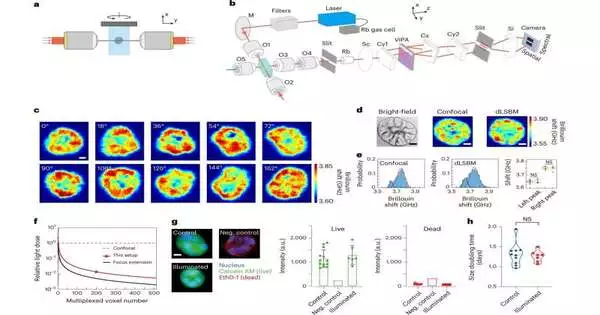
Zhang and his partners from the College of Maryland — where Zhang burned through six years in the Branch of Bioengineering prior to joining Wayne State in 2021 — and the National Institutes of Health (NIH) as of late distributed an examination article in Nature Strategies looking at the utilization of double line-filtering Brillouin microscopy (dLSBM) to further develop securing speed and diminish illumination dosages, two principal restricting variables to the far-reaching utilization of this procedure in biomedicine.
“Existing confocal Brillouin microscopy is genuinely sluggish; it requires a couple of moments to secure one mechanical picture of a solitary cell,” said Zhang. “On the off chance that we are imaging bigger examples, for example, growth cell bunches or beginning-phase undeveloped organisms, we really want to stand by an hour or longer to acquire one picture.”
Utilizing dLSBM, Zhang’s group announced paces of 50 to multiple times quicker than its partner, while lessening the light illumination level by multiple times for 2D and 3D mechanical planning.
“With this development, we can obtain a mechanical picture of cell bunches shortly,” he said. “This better-securing speed is significant in light of the fact that it permits us to explore subtleties of cell ways of behaving in practically constant.”
Brillouin microscopy is an optical imaging methodology established in what is known as Brillouin light dissipating (BLS), first revealed in 1922 by French physicist Léon Brillouin. BLS happens when light collaborates with a substance and warm changes or vibrations of particles in the material cause the light to dissipate. Vibrations can be impacted by specific variables, including heat, pressure, water content, or material solidness. The last option of these qualities is generally significant for the utilization of Brillouin microscopy as a symptomatic device.
Sickness movement, like malignant growth metastasis, is frequently connected with changes in cell firmness; however, this is challenging to gauge since cells are little and live in extremely delicate tissue. Traditional strategies measure arranged cells on a petri dish or other hard substrate. A Brillouin magnifying lens just uses a laser pillar to test the mechanical properties, permitting estimation to be led when cells are in their physiological circumstances.
Since actual contact isn’t required, brilliance in innovation is undeniably not so much obtrusive but rather more advantageous. One more application for which these attributes are significant is to more readily figure out early-stage tissue improvement, especially as it relates to birth sicknesses and problems.
“Because of the 3D design of an undeveloped organism, customary contact-based strategies experience enormous difficulties for in vivo estimation,” said Zhang. “Since Brillouin microscopy works in a non-contact way, it in some cases turns into the main accessible decision.”
Zhang teams up with scholars and specialists at Karmanos and different establishments to resolve biomedical inquiries with mechanical developments. Nonetheless, that’s what Zhang noticed: “Brillouin innovation is still in its beginning phase and has restricted imaging profundity. Our lab will keep on chipping away at making it more open for more extensive biomedical fields.”
The connection among specialists and individuals from the clinical local area is particularly important at the analytic phase of the medical services venture. Zhang and other Wayne State BME scientists are directing biomedical exploration and medical care to levels of unrivaled movement.
More information: Jitao Zhang et al, Rapid biomechanical imaging at low irradiation level via dual line-scanning Brillouin microscopy, Nature Methods (2023). DOI: 10.1038/s41592-023-01816-z


