A group of scientists affiliated with a few establishments in the U.S. has recognized a subtype of synapses that kick the bucket in Parkinson’s patients. In their review, distributed in the journal Nature Neuroscience, the group utilized another RNA sequencing method to investigate synapses in the substantia nigra and afterward thought about specific sorts they found in the minds of Parkinson’s patients and unafflicted subjects to recognize contrasts. Ernest Arenas, with the Karolinska Institutet in Sweden, distributed a News and Views piece in a similar diary issue illustrating how single-cell investigation of synapses is directed and remarking on the work announced by the group on this new effort.
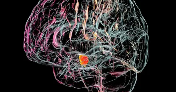
Parkinson’s infection is an ever-evolving neurodegenerative sickness — patients experience balance issues, inconvenience talking and quakes. There is no fix, but a few medications can lessen side effects. Earlier examination has shown that the infection emerges when nerve cells in the substantia nigra (situated in the midbrain) disintegrate for obscure reasons. Thus, less dopamine is created. As a greater number of the cells stop working, the side effects deteriorate. In this new study, the specialists investigated the nerve cells in the substantia nigra to figure out which of them bite the dust in Parkinson’s patients.
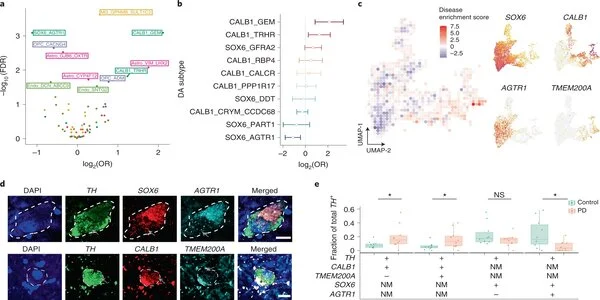
The researchers used a recently developed single-cell RNA method that sequences individual cells in a given tissue test. The scientists utilized it to figure out which qualities in cells in the substantia nigra were delivering proteins and afterward arranged them into 10 subtypes.
They then acquired memory tests from 10 individuals who had Parkinson’s (or Lewy body dementia) at the hour of their passing. They carried out a similar kind of RNA sequencing on the examples in general and, furthermore, on various mind tests gathered from unafflicted individuals after death. They thought about examples from the two gatherings searching for contrasts and found one of the diminished synapse subtypes in the Parkinson’s patients, recommending firmly that it is the most impacted in individuals with Parkinson’s.
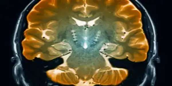
While checking for Parkinson’s illness, SPECT imaging of the cerebrum is utilized for procuring data on dopamine action. Another review directed in Turku, Finland, shows that the dopamine action seen in SPECT imaging doesn’t mirror the quantity of dopamine neurons in the substantia nigra, as recently expected.
One of the main changes in the focal sensory system in Parkinson’s sickness is the deficiency of dopamine-creating neurons in the substantia nigra, causing a drop in dopamine levels in the cerebrum.
For example, “Low dopamine level in the mind is connected with the focal engine side effects of Parkinson’s illness, for example, quaking or shaking, muscle solidity, and gradualness of development,” explains Docent of Neurology Valtteri Kaasinen from the University of Turku.
“Low dopamine level in the brain is linked with the central motor symptoms of Parkinson’s disease, i.e. tremor or shaking, muscle stiffness and slowness of movements,”
Docent of Neurology Valtteri Kaasinen from the University of Turku
Solitary photon outflow processed tomography (SPECT) imaging of the mind can identify diminished dopamine action. This technique is broadly utilized in the diagnostics of Parkinson’s infection in Europe and the United States.
The review led at the University of Turku and Turku University Hospital shows that the dopamine movement seen in SPECT imaging doesn’t mirror the quantity of dopamine neurons in the substantia nigra, in spite of what has been thought process. As indicated by Kaasinen, this is a significant outcome as it demonstrates that the relationship between the quantity of neurons and dopamine action isn’t direct.
“This should be considered later on while creating medicines that influence the quantity of neurons in the substantia nigra.” “It additionally appears to be that SPECT imaging is certainly not an appropriate strategy for checking whether treatment research brings about cutting-edge Parkinson’s infection while concentrating on medicines that influence the quantity of neurons in the substantia nigra,” says Kaasinen.
In the review, posthumous neuron numbers in the substantia nigra were determined for patients with Parkinson’s illness who had been analyzed with dopamine carrier SPECT before death. The quantity of neurons can’t be determined during a patient’s lifetime since the substantia nigra is found profound inside the midbrain where biopsy is unimaginable in vivo.


